Brain anatomy can be daunting. Let’s try to tackle it step by step and start with the surfaces of the cerebral hemispheres. The surface of the cerebral hemispheres consists of many convolutions , the cortical gyri, which are separated by sulci of varying sizes. Even if you aced neuroanatomy during medical school and remember all cortical gyri and sulci by heart (which you probably don’t :)), trying to find and recognize them on an MRI or CT-exam of the brain can seem like an impossible task at first. No worries, it isn’t. With a little bit of practice and some tips and tricks, you’ll be able to recognize the most important surface landmarks of the brain in no time. Let’s start with the simplest and most important one: the central sulcus.
The central sulcus is an important anatomic landmark on imaging. The central sulcus separates the frontal from the parietal lobe and is the sulcus between the precentral and the postcentralis gyrus, which constitute the primary motor cortex and primary somatosensory cortex respectively. The primary motor cortex is responsible for the control and execution of voluntary movements and lesions in the precentral gyrus will lead to a contralateral hemiparesis. The primary somatosensory cortex is responsible for the primary processing of somatic sensations.
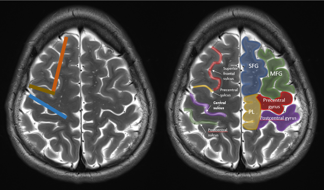
It’s easiest to find the central sulcus in the axial plane. I’ve chosen an axial T2-weighted image for this tutorial, but you can find the central sulcus on any sequence off course. The central sulcus is easiest to detect relatively high up in the brain, above the level of the lateral ventricles.

Let’s start with the easiest one (in my opinion, almost never lets me down). Do you see the deep sulcus anteriorly in the brain? That’s the superior frontal sulcus. I’ve coloured it red on the image below. The superior frontal sulcus separates the superior frontal gyrus (blue) from the middle frontal gyrus (green). At the posterior border of the middle frontal gyrus, the superior frontal sulcus (red) forms a juntion with the precentral sulcus (yellow). The combination of the superior frontal sulcus and the precentral sulcus looks a bit like capital letter “L” (or an inverted capital letter “L” depending on which hemisphere you’re looking at).

We’re almost there! The gyrus behind the precentral sulcus is the precentral gyrus (red). The sulcus behind the precentral gyrus is the central sulcus (purple), the gyrus behind that the postcentral gyrus (purple), and the sulcus behind that the postcentral sulcus (green). Easy, no? We’ve found the central sulcus and it took us no effort at all. Just look for capital letter “L” and the central sulcus is the sulcus immediately behind the foot of letter “L”.

Some small details: notice that the central sulcus doesn’t run all the way up to the medial hemispheric surface (sometimes it does, but most of the time it doesn’t). As a consequence, there’s not sulcus separating the pre- and postcentral gyrus at the medial surface of the brain. We call this part of the pericentral region “the paracentral lobule” and I’ve drawn it in yellow on the image above. The primary motor and somatosensory cortex are somatotopically organized and the paracentral lobule contains the motor (anterior) and somatosensory (posterior) representations of the contralateral leg. In other words: a hemorrhage or an infarction in the paracentral lobule, especially when anteriorly in the paracentral lobule, will cause a paresis of the contralateral leg. Also notice on the images above that in the left hemisphere, there’s some kind of “cortical bridge” between the middle frontal gyrus and the precentral gyrus, with no precentral sulcus separating the two gyri. This is just one of many possible anatomic variations we might encounter when studying brain surface anatomy, and that’s why we need some other tricks to recognize the central sulcus.
Finding the hand knob is another way to quickly detect the precntral gyrus. The motor representations in the primary motor cortex are somatotopically organized. The motor respresentation of the contralateral hand and arm are typically located in a dorsally oriented small “knob” on the upper end of the precentral gyrus. This is the so-called “hand knob” and in imaging studies the configuration of the hand knob on the precentral gyrus is sometimes reffered to as the “inverted omega sign” (in red on the image below). When you combine this with the “L-sign”, you are twice as sure that you’ve found the precentral gyrus!

But when it comes to the hand knob, a lot of anatomic variations are possible as well. Sometimes we see a clear “knob”, sometimes we see two knobs, sometimes we see no knob at all.
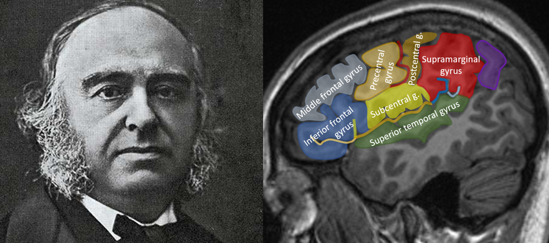
Let’s learn another trick that will help us find the precentral gyrus. For our last trick, we need to find the “marginal branch of the cingulate sulcus” in the axial plane. The cingulate sulcus is easily recognizable in the sagittale plane and is the paramedian sulcus on top the cingulate gyrus. At its posterior end, the sulcus runs upwards and towards the medial hemispheric surface and this is called the “marginal branch (of the cingulate sulcus)”. The image below shows the cingulate sulcus and it’s marginal branch in the sagittal plane.

In the axial plane, the marginal branch is (sometimes) easily recognizable because it forms a “bracket” on the medial brain surface with its contralateral brother or sister (and that’s why some call this the “pars bracket sign”). The central sulcus is located anterolateral of the “pars bracket”.

So let’s summarize: when combining the L-sign, the handknob-sign and the pars-bracket sign, we should be able to easily recognize the central sulcus in the axial plane on imaging. There are more tricks, but these are the quickest and easiest to use. Start to apply them on every brain scan you see, and after a while you’ll detect the central sulcus and precentral gyrus almost automatically without having to think too much about it. Try it and let me know how it worked out!
Please share your own experiences or additional tips and tricks! Looking forward to your comments and suggestions.
Hello could you please do a video on the cranial nerve anatomy and their course and pathology.
Thanks
Very nicely explained!
brilliant !
Cool and informative, studed these signs in text book but was unable able to fully understand.
Thanks!
Vero good
will there be any updates to this blog?
Yes, there, just been really busy with real life, but the blog ain’t dead 🙂
Yes, please post more!!!
Dan, neurology resident
The article was interesting and informative.
As clear as can ever be ! Thanks !
Thank you a lot. You really help us neurosurgeons as well
is there a key to the abbreviations?
Thank you so much
interesting is the central sulcus is not central